Transesophageal Echocardiography (TEE) Simulators
Advanced TEE simulation technology designed to train cardiologists, anesthesiologists, and cardiac sonographers in complex cardiac imaging procedures with zero risk to patients.

Advanced Cardiac Imaging
High-fidelity simulation of complex cardiac anatomy and pathologies.
Realistic Haptic Feedback
Authentic probe manipulation with realistic resistance and tactile feedback.
Comprehensive Case Library
Extensive collection of normal and pathological cardiac conditions.
Performance Analytics
Detailed metrics on image acquisition, interpretation, and procedural skills.
What is Transesophageal Echocardiography (TEE)?
Transesophageal Echocardiography (TEE) is an advanced cardiac imaging technique where an ultrasound probe is inserted through the esophagus to obtain high-resolution images of the heart. This procedure provides superior visualization of cardiac structures compared to transthoracic echocardiography, particularly for posterior cardiac structures like the left atrium, mitral valve, and thoracic aorta.
TEE is essential for diagnosing complex cardiac conditions, guiding interventional procedures, and monitoring cardiac function during surgery. However, it requires specialized training due to its invasive nature and the complexity of image interpretation. TEE simulators provide a safe, controlled environment for developing these critical skills without patient risk.
Intraoperative Monitoring
Real-time cardiac assessment during cardiac and non-cardiac surgery.
Valvular Assessment
Detailed evaluation of valve morphology, function, and pathology.
Congenital Heart Disease
Assessment of complex congenital cardiac anomalies and repairs.
Source of Embolism
Identification of cardiac sources of embolism in stroke patients.
Available TEE Simulators
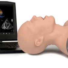
TEE Probe Trainer
Practice realistic probe insertion, manipulation, and positioning with haptic feedback.
Request Quote
Interactive TEE Case Scenarios
Comprehensive library of cardiac pathologies with guided diagnostic challenges.
Request Quote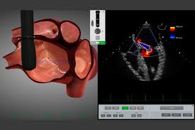
TEE Simulation Software
Advanced software with real-time image processing, measurements, and guided tutorials.
Request Quote
3D TEE Anatomy Trainer
Interactive 3D cardiac models for spatial understanding of cardiac structures.
Request Quote
Interventional TEE Simulator
Training for TEE-guided procedures including valve interventions and closures.
Request Quote
Emergency TEE Trainer
Rapid assessment training for critical care and emergency settings.
Request Quote
Pediatric TEE Simulator
Specialized training for congenital heart disease and pediatric cardiac assessment.
Request Quote
Comprehensive TEE Training Suite
Complete system with all TEE training modules and assessment tools.
Request QuoteComprehensive TEE Education and Training
Transesophageal Echocardiography (TEE) is a critical skill for cardiologists, cardiac anesthesiologists, and intensivists. TEE simulation provides a safe, controlled environment for learners to develop the complex psychomotor and cognitive skills required for this invasive procedure. Our TEE simulators bridge the gap between theoretical knowledge and clinical competence, ensuring healthcare professionals are fully prepared for real-world applications.
Our TEE training systems support development of:
- Probe insertion and manipulation techniques
- Standard TEE views and imaging planes
- Cardiac anatomy recognition and spatial orientation
- Pathology identification and differential diagnosis
- Quantitative measurements and calculations
- Doppler and color flow assessment
- 3D image acquisition and interpretation
- Intraoperative monitoring and guidance
TEE simulation is particularly valuable for:
- Cardiology Fellows: Developing expertise in diagnostic and interventional TEE
- Cardiac Anesthesiologists: Mastering intraoperative TEE for surgical guidance
- Cardiac Sonographers: Refining imaging techniques and pathology recognition
- Intensivists: Learning critical care TEE for hemodynamic assessment
- Cardiac Surgeons: Understanding TEE imaging for surgical planning
- Emergency Physicians: Training in emergent TEE for unstable patients
Our TEE simulators feature realistic haptic feedback, authentic console interfaces, and extensive case libraries that include normal anatomy, common pathologies, and complex cardiac conditions. The systems provide immediate feedback on probe manipulation, image acquisition, and interpretation skills, allowing learners to identify areas for improvement and track their progress over time.
TEE vs. Transthoracic Echocardiography (TTE)
| Feature | TEE | TTE |
|---|---|---|
| Image Quality | Superior (closer to heart, higher frequency) | Good (through chest wall) |
| Cardiac Structures | Excellent for posterior structures (LA, mitral valve) | Good for anterior structures (LV, RV) |
| Patient Preparation | Fasting, sedation, monitoring required | Minimal preparation needed |
| Procedure Time | 15-30 minutes (plus recovery) | 15-30 minutes |
| Risk Level | Low risk (sedation, esophageal insertion) | Very low risk (non-invasive) |
| Training Complexity | High (invasive procedure, complex interpretation) | Moderate (non-invasive, standard views) |